

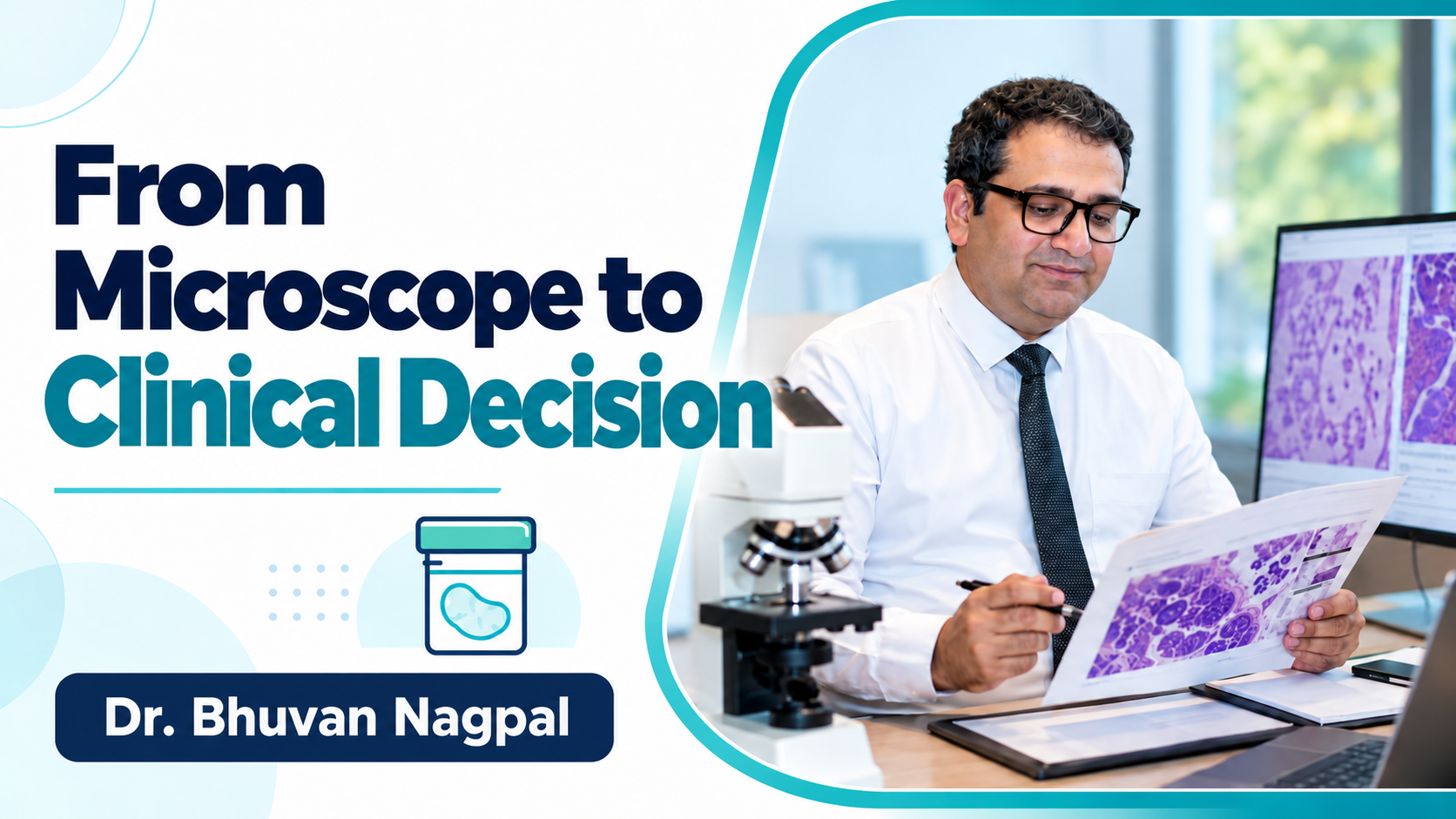
Diagnosis is a reasoned interpretation
Microscopy is central to tissue diagnosis, but the result is not simply the recognition of one familiar image. A pathology report brings together the examination of the specimen and relevant clinical information; additional studies may contribute when needed. [1]
For learners, it is helpful to think of the process as answering a series of questions while checking that the available material supports each answer.
Describe before naming
My suggested learning approach begins with an organised description. Identify what tissue is present, how it is arranged and which features seem most important. Then develop a short differential diagnosis and identify what would distinguish the possibilities.
This method helps avoid forcing every feature into the first diagnosis that comes to mind. It also makes a discussion with a supervisor more informative than presenting only a label.
Bring the clinical question back into view
Oral and maxillofacial pathology draws on clinical, radiographic and microscopic investigation. [2] Ask whether the proposed interpretation explains the lesion’s location and the findings supplied by the clinician.
If important information is missing, request it. If the tissue and clinical findings do not fit, identify the mismatch and discuss how to resolve it.
Use additional tests purposefully
Before requesting another investigation, state the question it should answer. After receiving the result, reassess the whole interpretation rather than simply adding another line to the report.
Communicate uncertainty with appropriate specificity. Explain whether the limitation concerns the sample, the information supplied or the ability to distinguish particular possibilities.
Make the report useful to the next decision
The treating clinician needs to understand what has been established and what, if anything, remains unresolved. Patients need a plain-language explanation of how the result affects their plan.
A good educational exercise is to ask: “What can the treating team reasonably do with this report?” That question encourages precise diagnosis, clear limitations and direct communication. The value of microscopy is realised when its findings contribute to an appropriate clinical decision.
References
[1] National Cancer Institute. Surgical Pathology Reports. Read source[2] American Academy of Oral and Maxillofacial Pathology. About AAOMP. Read source